Our eosinophilosophy on
WHY EOS COUNTS

Eosinophils are multifunctional leukocytes that can be both helpful and harmful, making their role difficult to determine.1,2 While eosinophil involvement in inflammatory disease is well-established, more is being understood about their role in health and how they can help guide diagnosis and treatment decisions.1,3-10

EOS counts in health

The role of eosinophils in health was long thought to be limited to defense against parasitic infections. It is now known that eosinophils could contribute to overall health and immunoregulation.2,11

Contribution to overall health and immunoregulation comes from both blood- and tissue-resident eosinophils

In fact, many blood- and tissue-resident eosinophils deliver protective effects in response to infections and disease. In addition to protection against parasitic infections, eosinophils are thought to play a role in host defense of some viral, bacterial, and fungal infections.11,12 Not only do eosinophils directly participate in innate immunity, they may have many roles in adaptive immunity, including regulation of T-cell development in the thymus, lymphocyte recruitment, antigen presentation, and driving T helper 2 (Th2) cell polarization.13 Resident eosinophils in the gastrointestinal tract have been shown to contribute to intestinal immune homeostasis by regulating T-cell and immunoglobulin A responses.14,15

Tissue-resident eosinophils are involved in tissue maintenance, metabolism, and immune homeostasis; however, their specific function depends on the tissue in which they reside.12,14 Under physiologic conditions, tissue-resident eosinophils have been identified in14:

Adipose tissue icon

Adipose tissue

Gastrointestinal tract icon

Gastrointestinal tract

Pulmonary icon

Lung

Mammary gland icon

Mammary gland

Spleen icon

Spleen

Uterus icon

Uterus

Eosinophils may also play a role in tissue repair through secretion of fibroblast growth factor and transforming growth factor-β. These growth factors are secreted from the eosinophil following epithelial damage.13

What is an appropriate blood EOS level?

107 Eosinophils/µL*

A recent study found mean blood eosinophil levels in a healthy population (n=3641) to be 107 eosinophils/μL. The same study found blood eosinophil counts are consistently higher in males (120 cells/μL median) than females (100 cells/μL median), are highest in infancy and adolescence, and are independent of age in adults. Additionally, atopy, smoking, asthma, chronic obstructive pulmonary disease, obesity, and metabolic syndrome can be associated with higher eosinophil counts.16*

*Geometric mean; 95% confidence interval (105-110 eosinophils/μL).

Although eosinophils may contribute to overall health, the impact of levels
below what is considered normal is not known11

EOS counts in disease

During immune responses mediated by Th2 cells and group 2 innate lymphoid cells (ILC2), eosinophils become activated and multiply. Th2 cells and ILC2 cells are major sources of interleukin 5 (IL-5), the main cytokine responsible for maturation, activation, and survival of eosinophils.12,17 Eosinophils release cytokines and chemokines to target foreign antigens, promote inflammation, and damage surrounding structures.1,12,13

Cytokines and chemokines released from eosinophils lead to inflammation and tissue damage12,17,18
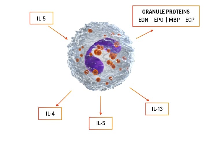
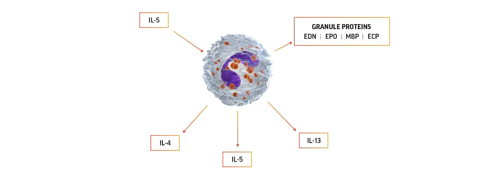
Eosinophil activation diagram

Eosinophil activation can lead to production of IL-5, which can promote
eosinophils’ own survival by eosinophilopoiesis and activation.

The 4 major proteins released from eosinophil granules and their functions include12:

Eosinophil cationic protein (ECP)

  • Causes membrane disruption
  • Cytotoxic to host cells and pathogens
  • Neurotoxic

Eosinophil-derived neurotoxin (EDN)

  • Antiviral
  • Neurotoxic
  • Maturation, activation, and chemotaxis of dendritic cells

Eosinophil peroxidase (EPO)

  • Leads to generation of reactive oxygen species, which are toxic to extracellular pathogens
  • Inflammatory effects
  • Anti-inflammatory effects

Major basic protein (MBP)

  • Causes membrane disruption
  • Cytotoxic to host cells and pathogens
  • Activates mast cells, basophils, and neutrophils
  • Neuroprotective effects

Reconsider the role of IL-5 in eosinophilic disease. Watch the video below to learn more about the impact of IL-5, eosinophils, and other inflammatory cells in type 2 inflammation.

  • TRANSCRIPT

    ONSCREEN TEXT:
    ReThink IL-5
    For US healthcare professionals only.
    NP-US-AAP-VID-240001, June 2024.

    Scene 1

    NARRATOR:
    Type 2 inflammation is the basis of pathophysiology for many inflammatory diseases.

    ONSCREEN TEXT:
    Type 2 inflammation is the basis of pathophysiology for many inflammatory diseases.1,2,3

    NARRATOR:
    Some of this inflammation is driven by interleukin-5, or IL-5, a powerful pro-inflammatory cytokine…

    ONSCREEN TEXT:
    Some of this inflammation is driven by interleukin-5, or IL-5, a powerful pro-inflammatory cytokine…

    NARRATOR:
    …which has been known for its role in the growth, differentiation, recruitment, activation, and survival of eosinophils.

    ONSCREEN TEXT:
    …which has been known for its role in the growth, differentiation, recruitment, activation, and survival of eosinophils.2,4

    NARRATOR:
    Increased levels of IL-5 are responsible for the elevated levels of eosinophils seen in patients with diseases like asthma with T2 inflammation. Activated eosinophils exhibit cytotoxic activity, release cytokines, and modulate other immune cells.

    ON-SCREEN TEXT:
    Increased levels of IL-5 are responsible for the elevated levels of eosinophils seen in patients with diseases like asthma with T2 inflammation.2,5,6,7 Activated eosinophils exhibit cytotoxic activity, release cytokines, and modulate other immune cells.8

    NARRATOR:
    This contributes to epithelial barrier damage, tissue remodelling, and airway inflammation and makes eosinophils key to the pathophysiology of these conditions.

    ON-SCREEN TEXT:
    This contributes to epithelial barrier damage, tissue remodelling, and airway inflammation and makes eosinophils key to the pathophysiology of these conditions.5,8,9,10,11

    NARRATOR:
    Evolving science suggests it is time to rethink the role of IL-5 in Type 2 inflammation.

    ONSCREEN TEXT:
    Evolving science suggests it is time to rethink the role of IL-5 in Type 2 inflammation.

    Scene 2

    NARRATOR:
    IL-5 is a core cytokine that may orchestrate multidirectional effects on a range of cell types, including T cells, plasma cells, ILC2 cells, mast cells, fibroblasts, epithelial cells, and eosinophils.

    ON-SCREEN TEXT:
    IL-5 is a core cytokine that may orchestrate multidirectional effects on a range of cell types, including T cells, plasma cells, ILC2 cells, mast cells, fibroblasts, epithelial cells, and eosinophils.5,6,11,13,14,15,16,17,18

    NARRATOR:
    These cells are responsible for inflammation.

    ON-SCREEN TEXT:
    These cells are responsible for inflammation.5,7,11,13,14,15,16,17,18

    NARRATOR:
    For example, IL-5 has been shown to impact key cellular targets of Type 2 inflammation…

    ON-SCREEN TEXT:
    For example, IL-5 has been shown to impact key cellular targets of Type 2 inflammation…

    NARRATOR:
    …with IL-5 receptors being present and active on mast cells and plasma cells.

    ONSCREEN TEXT:
    …with IL-5 receptors being present and active on mast cells and plasma cells.17,18

    NARRATOR:
    Furthermore, IL-5 appears to affect both T cell and ILC2 cell activity.

    ON-SCREEN TEXT:
    Furthermore, IL-5 appears to affect both T cell and ILC2 cell activity.16,19

    Scene 3

    NARRATOR:
    Epithelial cells and fibroblasts both play a key role in regulating epithelial barrier function and tissue repair.

    ONSCREEN TEXT:
    Epithelial cells and fibroblasts both play a key role in regulating epithelial barrier function and tissue repair.13,14

    NARRATOR:
    IL-5 receptors were found to be present and active on the surface of epithelial cells and fibroblasts.

    ONSCREEN TEXT:
    IL-5 receptors were found to be present and active on the surface of epithelial cells and fibroblasts.13,14,18

    NARRATOR:
    Increased IL-5 may promote epithelial barrier dysfunction and tissue fibrosis through cellular effects on tight junctions, the basement membrane, and collagen production.

    ONSCREEN TEXT:
    Increased IL-5 may promote epithelial barrier dysfunction and tissue fibrosis through cellular effects on tight junctions, the basement membrane, and collagen production.13,14,18

    Scene 4

    NARRATOR:
    Impaired mucus secretion has been associated with IL-5-mediated effects on several cells, including eosinophils.

    ONSCREEN TEXT:
    Impaired mucus secretion has been associated with IL-5-mediated effects on several cells, including eosinophils.7,15,16,17,18,20,21

    NARRATOR:
    In summary, this emerging science suggests that IL-5 has a role in inflammatory changes, including airway remodeling, epithelial barrier dysfunction, and mucus plugging, contributing to a more holistic understanding of IL-5 pathophysiology in the Type 2 inflammatory process.

    ONSCREEN TEXT:
    In summary, this emerging science suggests that IL-5 has a role in inflammatory changes, including airway remodeling, epithelial barrier dysfunction, and mucus plugging, contributing to a more holistic understanding of IL-5 pathophysiology in the Type 2 inflammatory process.7,13,14,15,16,17,18,20,21,22

    Outro
    GSK logo

    REFERENCES:
    1. Senna G et al. Multidiscip Respir Med. 2022;17(1):318.
    2. Pelaia C et al. Front Physiol. 2019;10:1514.
    3. Brusselle G, Bracke K. Ann Am Thorac Soc. 2014;11 Suppl 5:S322-S328.
    4. Park YM, Bochner BS. Allergy Asthma Immunol Res. 2010;2(2):87-101.
    5. Gevaert P et al. Int Forum Allergy Rhinol. 2022;12(11):1413-1423.
    6. Tomassen P et al. J Allergy Clin Immonul. 2016;137(5):1449-1456.e4.
    7. Pelaia G et al. Mediators Inflamm. 2015;2015:879783.
    8. Varricchi G et al. Curr Opin Allergy Clin Immunol. 2016;16(2):186-200.
    9. Naik SR, Wala SM. Recent Pat Inflamm Allergy Drug Discov. 2013;7(1):62-95.
    10. Tosun F et al. Ann Otol Rhinol Laryngol. 2010;94(1):e13031.
    11. Vaglio A et al. Allergy. 2013;68(3):261-273.
    12. Valent P et al. J Allergy Clin Immunol. 2012;130(3):707-612.e9.
    13. Barretto KT et al. Allergy. 2020;75(8)2127-2130.
    14. Bajbouj K et al. Allergy. 2023;78(3)882-885.
    15. Bergantini L et al. Scand J Immonul. 2021;94(1):e13031.
    16. Malik B et al. Respirology. 2023;28(8):758-766.
    17. Buchheit KM et al. J Allergy Clin Immunol. 2020;145(6):1574-1584.
    18. Buchheit KM et al. J Allergy Clin Immunol. 2021;148(2):574-584.
    19. Bergantini L et al. Biomed Pharmacother. 2023;11(1)195-199.e2.
    20. Dunican EM et al. J Clin Invest. 2018 ;128(3):997-1009.
    21. Chan R et al. J Allergy Clin Immunol Pract. 2023 ;11(1):195-199.e2.
    22. Huang Y, Qiu C. Ann Transl Med. 2022;10(18):1023.

    Trademarks are owned by or licensed to the GSK group of companies.
    ©2024 GSK or licensor.
    NP-US-AAP-VID-240001, June 2024
    Produced in USA.

EOS counts in diagnosis and treatment decisions

Blood and/or tissue eosinophils can be used as a biomarker in clinical practice to guide diagnosis and treatment. Your patients’ blood eosinophil levels can be measured through a complete blood count with differential.3-10

Get the latest eosinophilosophy updates

References:

  1. Weller PF, Spencer LA. Functions of tissue-resident eosinophils. Nat Rev Immunol. 2017;17(12):746-760. 
  2. Travers J, Rothenberg ME. Eosinophils in mucosal immune responses. Mucosal Immunol. 2015;8(3):464-475. 
  3. Global Initiative for Asthma (GINA). Global strategy for asthma management and prevention. 2024 main report. https://ginasthma.org/2024-report/. Accessed July 25, 2024.
  4. Global Initiative for Chronic Obstructive Lung Disease (GOLD). Global strategy for the diagnosis, management, and prevention of chronic obstructive pulmonary disease. 2024 report. https://goldcopd.org/2024-gold-report/. Accessed July 25, 2024.
  5. Asano K, Hebisawa A, Ishiguro T, et al. New clinical diagnostic criteria for allergic bronchopulmonary aspergillosis/mycosis and its validation. J Allergy Clin Immunol. 2021;147(4):1261-1268.e5.
  6. Wechsler ME. Pulmonary eosinophilic syndromes. Immunol Allergy Clin North Am. 2007;27(3):477-492.
  7. Suzuki Y, Suda T. Eosinophilic pneumonia: a review of the previous literature, causes, diagnosis, and management. Allergol Int. 2019;68(4):413-419.
  8. Masi AT, Hunder GG, Lie JT, et al. The American College of Rheumatology 1990 criteria for the classification of Churg-Strauss syndrome (allergic granulomatosis and angiitis). Arthritis Rheum. 1990;33(8):1094-1100.
  9. Reed CC, Dellon ES. Eosinophilic esophagitis. Med Clin North Am. 2019;103(1):29-42.
  10. Valent P, Klion AD, Horny HP, et al. Contemporary consensus proposal on criteria and classification of eosinophilic disorders and related syndromes. J Allergy Clin Immunol. 2012;130(3):607-612.e9.
  11. Klion AD, Ackerman SJ, Bochner BS. Contributions of eosinophils to human health and disease. Annu Rev Pathol. 2020;15:179-209.
  12. Ramirez GA, Yacoub MR, Ripa M, et al. Eosinophils from physiology to disease: a comprehensive review. Biomed Res Int. 2018;2018:9095275.
  13. Wen T, Rothenberg ME. The regulatory function of eosinophils. Microbiol Spectr. 2016;4(5):10.1128/microbiolspec.MCHD-0020-2015.
  14. Marichal T, Mesnil C, Bureau F. Homeostatic eosinophils: characteristics and functions. Front Med (Lausanne). 2017;4:101.
  15. Chu DK, Jimenez-Saiz R, Verschoor CP, et al. Indigenous enteric eosinophils control DCs to initiate a primary Th2 immune response in vivo. J Exp Med. 2014;211(8):1657-1672.
  16. Hartl S, Breyer MK, Burghuber OC, et al. Blood eosinophil count in the general population: typical values and potential confounders. Eur Respir J. 2020;55(5):1901874.
  17. Kouro T, Takatsu K. IL-5- and eosinophil-mediated inflammation: from discovery to therapy. Int Immunol. 2009;21(12):1303-1309.
  18. Varricchi G, Senna G, Loffredo S, Bagnasco D, Ferrando M, Canonica GW. Reslizumab and eosinophilic asthma: one step closer to precision medicine? Front Immunol. 2017;8:242.